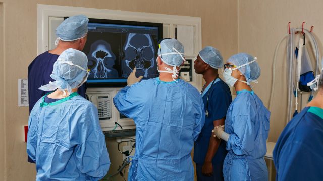
Interventional radiology: Study on occupational radiation exposure for medical staff
Compliance with occupational exposure limits
Each X-ray examination is associated with a certain radiation exposure to the patient. This is also the case for medical staff, especially during interventional procedures. The X-ray Ordinance and the Radiation Protection Ordinance lay down statutory exposure limits in order to protect the staff. Since different parts of the body react differently to radiation, there are dose limits for organs. Highly radiosensitive are, for example, bone marrow and gonads.
However, an explicit description of the occupational radiation exposure is a major challenge in interventional radiology. The reasons for this are the variety and multitude of applications as well as the different durations and the complexity of the procedures. In addition, the medical staff moves during the procedures and different body parts and organs are exposed to widely differing radiation fields. As a result, the so far collected measurement data on occupational exposure vary widely from study to study. Even within individual studies, the values deviate from one another for the same procedure.
Therefore, it is hardly possible to draw general conclusions about occupational radiation exposure so that a possible exceedance of limits for specific body parts might not be detected. The problem is partly attributable to the fact that specific body parts, such as hands or eyes, can be particularly highly exposed and, at the same time, are not adequately protected. With the classical dosimeters used, however, these partial body doses cannot be fully determined.
Measurement and calculation of radiation exposure in interventional radiology
Together with the Klinikum Augsburg, GRS contributes to improving the data situation with a new research project. “Setting up such research collaborations is an important element in the establishment of our university hospital”, says medical director Prof. Dr. Michael Beyer.
The objective of the project funded by the Federal Ministry for the Environment, Nature Conservation, Building and Nuclear Safety is the determination of occupational radiation exposure. On the one hand, this is done by measurements on site. For this purpose, typical situations are simulated in a so-called intervention room and radiation exposure is measured by means of dosimeters installed in the room. On the other hand, a simulation program is used for the calculation of radiation exposure. The simulation program is to be adapted and validated on the basis of the experimental measurements in order to reproduce the conditions as realistically as possible. Thus, the simulation program can also be used to calculate the radiation exposure of specific body part-regions which cannot be simply determined by dosimetry.
The project aims at improving occupational safety by providing a more accurate picture of radiation exposure in an intervention room. Changes or new measures to reduce radiation exposure, such as organisational recommendations or technical requirements, can then be assessed by simulation.
Benefits and risks of X-radiation
X-radiation is a form of ionising radiation. On its way through tissue the radiation deposits energy and might damage the genetic material of the cells. However, in contrast to the staff there are no dose limits for the patient but diagnostic reference levels which shall be taken as a basis for patient examinations. Furthermore, the X-ray Ordinance and the Radiation Protection Ordinance stipulate that each radiation application shall be medically justified and radiation exposure to the patient shall be kept as low as possible. This means, on the one hand, that the benefits and risks for the patient must be weighed up. On the other hand, the effect of radiation is to be controlled and reduced by means of specific safety precautions as far as it is compatible with medical requirements. For example, the radiation at specific body parts can be almost completely shielded by a lead shielding.
Radiation exposure varies depending on the used X-ray technique. A basic distinction is made between three techniques: conventional radiography, X-ray fluoroscopy and computed tomography (CT). The most commonly used technique is radiography. Here, radiation exposure to the patient is relatively small. The effective dose from a dental X-ray, for example, is about 0.01 mSv. For comparison, the average natural radiation exposure per person and year in Germany is about 2.2 mSv. On the other hand, a CT of the abdomen leads to a significantly higher effective dose for the patient.
Project highlights Radiation Protection

In the project, the GRS research team developed a central database for all information on water supply facilities in Germany that is relevant for radiation protection.